Advances in ultrasound technology help detect cancer better in women with dense breasts
The latest generation of breast cancer screening equipment has entered the Savannah market, and it boils down to a matter of black and white. Would you rather look for something tiny, dangerous and white on a white background, or something tiny, dangerous and black on a white background?
That’s an extremely simplified description of what a radiologist faces when reading the diagnostic scans of women with dense breast tissue. The mammogram leaves the physician trying to interpret white-on-white, while a breast ultrasound makes the picture far clearer. And the new equipment, the Automated Breast Ultrasound System (ABUS), takes the previous generation of hand-held breast ultrasound to the next level.
Read more: Breast Cancer by the Numbers: near 100% survival rate when caught in earliest stages
The October forecast?: Heightened awareness of breast cancer risks
“With ultrasound, cancer is black in a sea of white: instead of white-on-white, it’s black-on-white,” explained Dr. Jordan Dixon, D.O., who is the director of Telfair Women’s Imaging at Candler Hospital in Savannah.
The Telfair Breast Imaging Center at Candler Hospital acquired the automated equipment last December. The Telfair facility is the flagship breast facility for St. Joseph’s/Candler Health System, which also performs mammograms and handheld ultrasounds at St. Joseph’s and at the health system’s new campus in Pooler.
40 percent of women have dense breasts
So, what’s the difference between the handheld and the automated versions of the technology?
According to research published by National Institutes of Health’s Center for Biotechnology Information, ABUS performs scans faster, its scans are a quicker read for radiologists, and the scans are immensely more detailed since they generate literally thousands of images compared to the handful of views that usually result from the first-generation handheld version of the device.
“About 40 percent of patients have dense breasts, and when you do have dense breasts, you have a higher risk of developing breast cancer,” Dixon said.
Read more: St. Joseph’s/Candler honors Cheryl Rawlings as 2022 James R. Lientz Humanitarian
Given that, patients with dense breasts are usually advised to undergo a second, different screening with an ultrasound to pinpoint any possible abnormalities the mammogram might not have discovered.
“We have had patients who have dense breast tissue that have a normal mammogram and then had an ultrasound that was positive. Even in retrospect, you cannot see it on the mammogram,” Dixon said. “At some point it would have been large enough to find with mammography, but now we don’t have to wait.”
One thing a mammogram tells a woman is whether her breasts are dense or, like 60 percent of women, are more fatty in makeup. Dixon says a breast self-exam or a clinical exam conducted by a professional cannot distinguish between the two types. A woman’s weight or breast size is also no indication, because breast density is largely driven by genetics. But, once she has a mammogram, women in Georgia get official notification of whether their breasts are considered dense and how further screening is recommended. Since 2019, Georgia law has dictated that that information be included in the formal follow-up letter after a mammogram.
But even when women are armed with that information, many don’t heed the advice or don’t fully understand it. Dixon reports that Telfair does about six ABUS scans per day, while it performs some 120 mammograms on the average day. In that case, given the 40 percent of the population that has dense breasts, the six scans Telfair performs daily should be closer to 48.
Patients can pretty much assume that insurance will cover the cost of a mammogram, but ABUS is not so universally covered. However, Dixon said, her patients have generally found it to be covered.
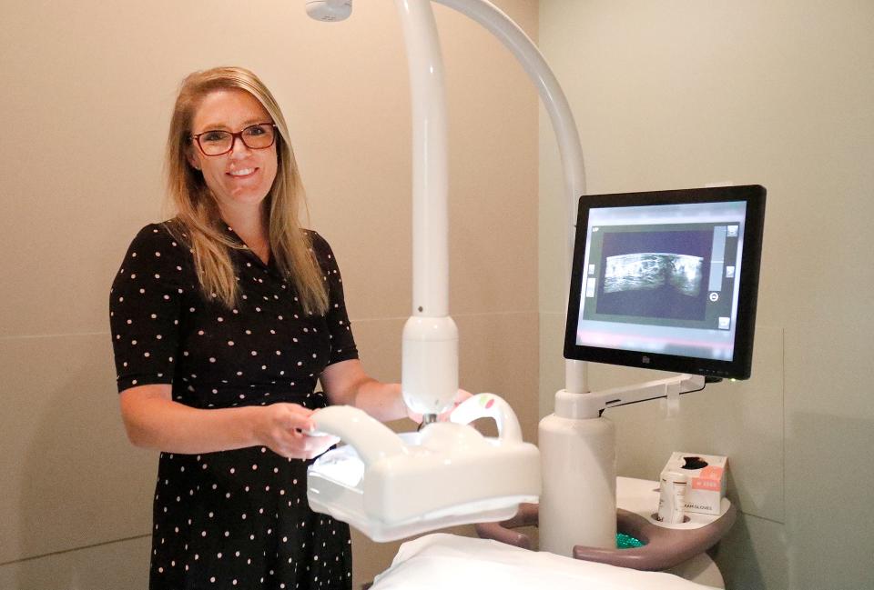
Dixon can describe the ABUS exam from personal experience.
“You’re lying on your back on a comfy bed with your arm above your head. They put lotion on breast and then they put this transducer over the breast, a large paddle, and that paddle is locked into place. It takes about 15 minutes per breast,” she said, and most patients get it done at the same appointment as their mammogram.
Mammograms have been a staple of women’s health for years – the American Cancer Society began recommending them back in 1976 – and despite the innovations to screening technology, they remain the gold standard for early detection of breast cancer. For women with dense breasts, their regular health regimen now may include ABUS and, for those considered at particularly high risk, breast MRI.
This article originally appeared on Savannah Morning News: Candler Hospital: Ultrasound advances help detect cancer in women

