Google's 3D Map of a Fly Brain Is Beautiful
Google and Janeila, a research campus at the Howard Hughes Medical Institute in Loudoun County, Virginia, have created "the most complete map of the fly brain ever created."
Their research was published earlier this week in the biology pre-print journal bioRxiv.
Connectomes, the maps illustrating connections in the brain, have been criticized by some scientists in the past because so far, they haven't led to any major breakthroughs.
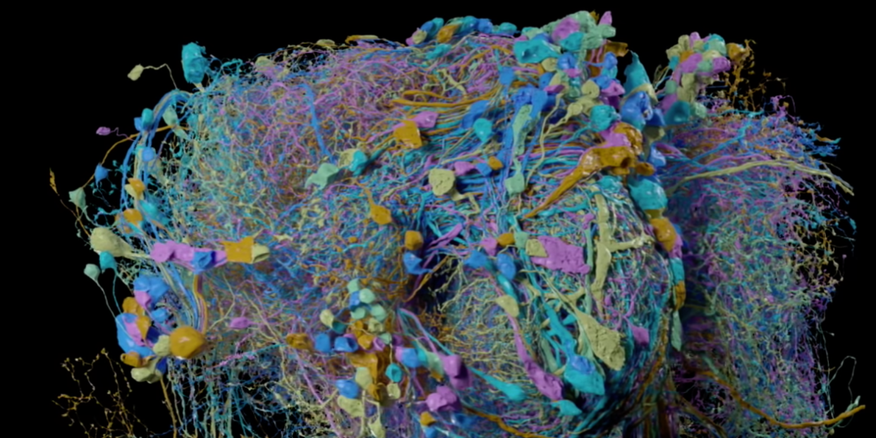
Drosophila melanogaster, otherwise known as the fruit fly, has a pretty impressive dome for being so small—as in, its brain contains over 100,000 neurons, or brain cells. For the first time, we can see about 25,000 of those neurons, across 4,000 different kinds, and their millions and millions of connections in the brain.
Google and Janeila Research Campus, part of the Howard Hughes Medical Institute in Loudoun County, Virginia, have created what they claim to be the world's largest and most detailed map of a brain. Called a wiring diagram, or a connectome, the map looks a bit like a 3D pile of Silly String, using neon colors to represent various portions of the brain that scientists are interested in studying. The researchers can even pull apart the map and isolate brain regions dealing with a fly's sense of smell, vision, or ability to navigate, drilling down into the brain connections they'd like to see.
"This was a big bet on something people thought was almost impossible to do," Viren Jain, a research scientist at Google and former laboratory head at Janelia, said in a press statement. "This will be the first time that we can really have a nuanced look at the organization of a nervous system with 100,000 neurons on a synaptic scale."
The researchers nicknamed the lucky insect in question, a female fruit fly, "hemibrain." Without high-powered microscopes, data analysis and advances in imaging tech, and deep learning algorithms, this project could have taken two decades, according to Gerry Rubin, vice president of Howard Hughes Medical Institute and executive director of Janeli.
Now the map can be used to study fly behavior by analyzing certain portions of the brain under certain conditions. The only question is whether or not critics who say connectomes aren't really useful will ever come around. In the past, these maps have faced pushback because they take so long to put together and haven't yet yielded any major breakthroughs.
A Brief History of Connectomes
To date, only one animal has ever had its full connectome published: Caenorhabditis elegans, a transparent roundworm that's only about one millimeter long. In the 1970s, a biologist named Sydney Brenner began preserving parts of the worm, cutting up thin slices of its body and taking photos of it under an electron microscope. By 1986, a near-complete version of the connectome was published, showing off almost all of the worm's 302 neurons and 7,000 connections—called synapses—between them.
The crazy thing? This was all done by hand. Two decades later, Dmitri Chklovskii, then a group leader at Janeila, finished the work by publishing a more detailed version of the worm connectome. At the time, it was the largest map of the brain to date.
Since then, scientists have done work to map out the brain of a rat, and of course, the fruit fly. Sebastian Seung, a noted scientist from the Princeton Neuroscience Institute, is an outspoken proponent of the connectome, even giving a TED Talk on the subject back in 2010.
Still, criticism remains. To create a connectome at the scale of the human brain—which contains about 86 billion neurons and 100 trillion synapses—would be a massive effort. In a 2012 debate with Seung, Anthony Movshon, a neuroscientist at New York University, pointed out he doesn't believe the C. elegans connectome has led to any major new insights about the worm's behavior:
I think it's fair to say…that our understanding of the worm has not been materially enhanced by having that connectome available to us. We don't have a comprehensive model of how the worm's nervous system actually produces the behaviors. What we have is a sort of a bed on which we can build experiments—and many people have built many elegant experiments on that bed. But that connectome by itself has not explained anything.
This appears to be one of those situations where the technology has somehow developed more quickly than the science, so it's hard to say whether or not these connectomes will be central to finding the next big truths about the brain.
Imaging the Fly Brain
To begin the mapping process, the researchers used a five-day-old female fruit fly. Then, they wrote in their paper, they used a custom jig to microdissect the fly's central nervous system, which they embedded in an epoxy resin called Epon. What resulted was a fully stained fly brain with synapses dyed even darker, which made it easier for a machine to detect those connections. The brain sections were then sliced into thin pieces at a thickness of just 20 microns each.
Using eight microscopes that were originally meant to capture data over a period of minutes or hours, the researchers configured them to run continuously for months or years. They used a technique called focused-ion beam scanning electron microscopy to capture the intricacies of the tiny brain's connections—it's only about the size of a poppy seed.
In this method, the microscope uses a focused beam of ions to gently blast away small pieces of brain tissue before it then shoots gallium ions at the tissue to polish it at the atomic level. This microscope takes an image of the tissue, polishes off another layer and then continues like that until the entire sample has been imaged. As the real fly brain is slowly shaved apart, a digital version forms.
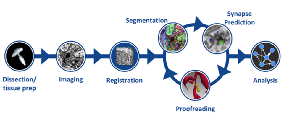
To store all of those images, it would take about 100 terabytes of space, or about the same amount of space your computer would need to keep 100 million photos. So researchers devised computer algorithms to stitch the images together, automating the process that was once done by hand to map the worm's brain.
The trick was teaching a computer how to break up images into smaller pieces, labeling each part. This process, called image segmentation, has been a focus of Google's for some time, but teaching an algorithm to look for neurons within microscope images takes a huge amount of training data. It's further complicated by the fact that neurons splay their tendrils across large portions of the brain, creating connections in many regions and ultimately spanning many images.
Eventually, the scientists ended up using what's called a flood-filling network, which could follow the neurons' tendrils from one end to the other as it snaked its way through the images. Based on prior experience, the algorithm can start making predictions about the shape of the tendrils and where they may lead.
The researchers estimated that Google's help in the computing sped up the process tenfold. Human proofreaders were still required, though, because the algorithms aren't perfect. "There’s still a lot of manual effort required," Ruchi Parekh, who leads a team of neuron tracers and proofreaders, said in a statement.
The researchers are now working on completing the full connectome while they continue to learn how to make sense of the images they've collected. Their goal? To learn something new about how the fly brain functions in the near future. Meanwhile, those heavy-duty microscopes are still hard at work, collecting images from a male fly's brain. This time, the researchers want to capture its entire nervous system—not just what's in the brain. If it all goes to plan, the images should all be scanned by the end of this year.
You Might Also Like

